


The discovery of X-rays is a fascinating tale that dates back to the late 19th century. In 1895, German physicist Wilhelm Conrad Röntgen stumbled upon this groundbreaking technology while experimenting with cathode rays. He noticed that a fluorescent screen in his lab began to glow even though it was not in direct contact with the cathode ray tube. Intrigued, Röntgen conducted further experiments and ultimately produced the first X-ray image of his wife’s hand, revealing her wedding ring. This moment marked the birth of a revolutionary diagnostic tool that would change the landscape of medicine forever.
Röntgen’s discovery quickly gained attention, and within a few years, X-rays were being used in medical settings across Europe and the United States. The initial excitement was palpable, as doctors began to realize the potential of X-rays for visualizing internal structures without invasive procedures. However, it wasn’t long before the dangers of radiation exposure became apparent. Early practitioners often suffered from radiation burns and other health issues due to inadequate safety measures. Despite these challenges, the medical community embraced X-rays, leading to rapid advancements in imaging techniques and equipment.
X-ray technology has revolutionized various fields, particularly in medical diagnostics and materials science. For those interested in enhancing their understanding of complex academic texts related to such technologies, an insightful resource is available. You can explore effective strategies for reading academic materials in the article titled “Strategies for Effective Reading of Academic Texts,” which can be found at this link. This article provides valuable tips that can aid in comprehending intricate subjects like X-ray applications and advancements.
Key Takeaways
- X-Rays were discovered by Wilhelm Conrad Roentgen in 1895, earning him the Nobel Prize in Physics in 1901.
- X-Rays work by passing electromagnetic radiation through the body to create an image on a film or digital detector.
- X-Rays are commonly used in medicine for diagnosing fractures, detecting tumors, and guiding medical procedures.
- Advantages of X-Rays include their ability to quickly produce images, while limitations include their potential for radiation exposure.
- Safety precautions for X-Ray procedures include wearing lead aprons and thyroid shields, and minimizing radiation exposure for pregnant patients.
How X-Rays Work
At its core, the process of generating X-rays involves the interaction of high-energy electrons with matter. When an X-ray machine is activated, electrons are accelerated and directed toward a metal target, typically made of tungsten. Upon striking this target, the electrons lose energy, which is emitted in the form of X-ray photons. These photons have the ability to penetrate various tissues in the body, depending on their density.
When X-rays pass through the body, they are absorbed at different rates by different tissues. Dense materials like bones absorb more X-rays and appear white on the resulting image, while softer tissues allow more X-rays to pass through and appear darker. This contrast creates a detailed image that can help healthcare professionals identify abnormalities or injuries. The entire process is quick, often taking just a few seconds, making it a convenient option for both patients and medical staff.
Uses of X-Rays in Medicine
X-rays have become an indispensable tool in modern medicine, serving a variety of purposes beyond just diagnosing fractures. One of the most common uses is in dental imaging, where X-rays help dentists identify cavities, gum disease, and other oral health issues that may not be visible during a routine examination. This early detection can lead to more effective treatment and better outcomes for patients.
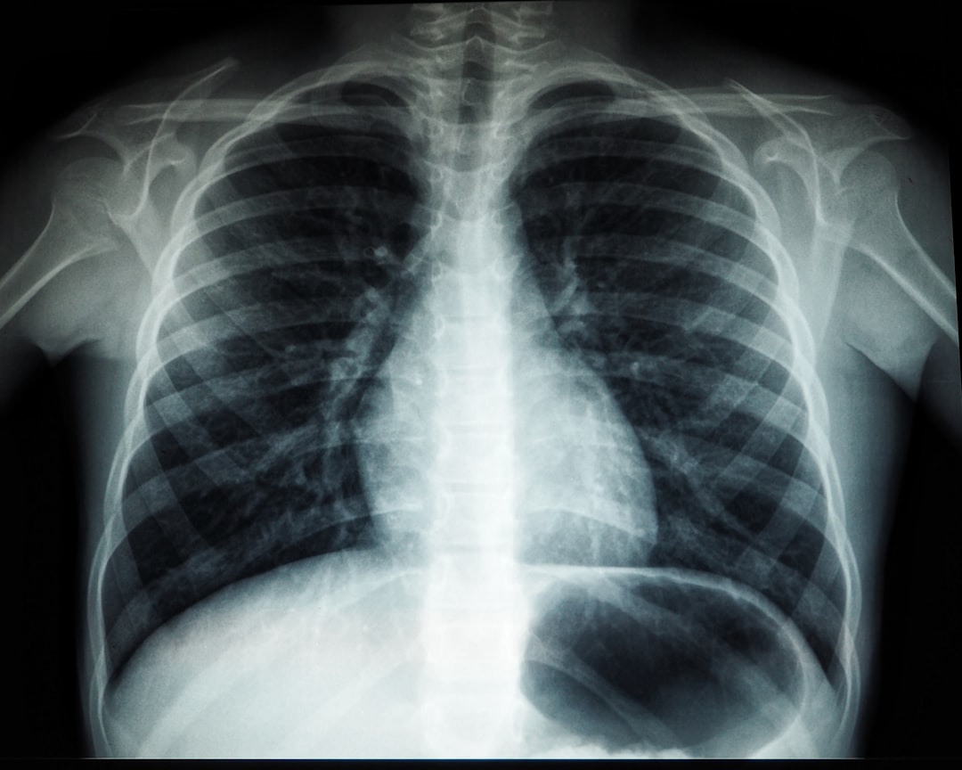
In addition to dental applications, X-rays are widely used in oncology to monitor tumors and assess treatment effectiveness. Radiologists can evaluate changes in tumor size or shape over time, providing crucial information for oncologists as they develop treatment plans. Furthermore, X-rays play a vital role in detecting conditions such as pneumonia or tuberculosis in the lungs, allowing for timely intervention and management of respiratory diseases.
Advantages and Limitations of X-Rays
One of the primary advantages of X-ray imaging is its speed and efficiency. The process is typically quick, allowing for rapid diagnosis and treatment decisions. Additionally, X-rays are relatively inexpensive compared to other imaging modalities like MRI or CT scans, making them accessible for many healthcare facilities.
Their ability to provide clear images of bone structures makes them particularly valuable in emergency settings where time is of the essence.
However, there are limitations to consider as well. One significant drawback is the exposure to ionizing radiation, which carries a risk of potential harm over time. While the amount of radiation from a single X-ray is generally low and considered safe for most patients, repeated exposure can accumulate and increase the risk of cancer. Moreover, X-rays are not as effective for visualizing soft tissues compared to other imaging techniques like MRI or ultrasound, which can limit their utility in certain diagnostic scenarios.
X-rays have revolutionized the field of medicine by allowing for non-invasive imaging of the human body, but their applications extend beyond healthcare. For those interested in the intersection of art and science, a fascinating article explores the philosophical underpinnings of Indian aesthetics and its multifaceted functions. This piece delves into how art can reflect deeper truths about existence, much like how X-rays reveal hidden structures within the body. To learn more about this intriguing relationship, you can read the article here.
Safety Precautions for X-Ray Procedures
| Category | Metrics |
|---|---|
| Usage | Number of X-ray scans performed |
| Accuracy | Percentage of accurate X-ray diagnoses |
| Cost | Average cost per X-ray scan |
| Technology | Types of X-ray machines used |
Given the risks associated with radiation exposure, safety precautions are essential during X-ray procedures. Healthcare providers typically follow strict protocols to minimize exposure for both patients and staff. For instance, lead aprons or shields are often used to protect sensitive areas of the body from unnecessary radiation during imaging.
Additionally, it’s crucial for patients to inform their healthcare providers about any previous X-ray procedures they’ve undergone, especially if they are pregnant or may be pregnant. In such cases, alternative imaging methods may be considered to avoid potential risks to the developing fetus.
Overall, maintaining open communication between patients and healthcare professionals is key to ensuring safety during X-ray examinations.
The Role of X-Rays in Disease Diagnosis
X-rays play a pivotal role in diagnosing various diseases and conditions across multiple medical specialties. In orthopedics, they are essential for identifying fractures, dislocations, and degenerative joint diseases like arthritis. By providing clear images of bone structures, X-rays enable orthopedic surgeons to make informed decisions regarding treatment options.
In addition to musculoskeletal issues, X-rays are crucial in detecting diseases affecting internal organs. For example, chest X-rays are commonly used to identify conditions such as pneumonia, heart failure, or lung cancer. By visualizing the lungs and surrounding structures, healthcare providers can assess the severity of these conditions and determine appropriate interventions. This diagnostic capability underscores the importance of X-rays in comprehensive patient care.
Advances in X-Ray Technology
Over the years, advancements in X-ray technology have significantly improved both image quality and patient safety. Digital radiography has largely replaced traditional film-based systems, allowing for faster image acquisition and enhanced diagnostic capabilities. Digital images can be manipulated for better visualization and can be easily shared among healthcare providers for collaborative decision-making.
Moreover, innovations such as computed tomography (CT) have expanded the applications of X-ray technology beyond traditional imaging. CT scans combine multiple X-ray images taken from different angles to create cross-sectional views of the body, providing detailed information about internal structures. This advancement has revolutionized diagnostics in various fields, including oncology and emergency medicine.
Future Applications of X-Rays
Looking ahead, the future applications of X-ray technology hold great promise for enhancing medical diagnostics and treatment options. One area of interest is the development of portable X-ray machines that can be used in remote or underserved areas where access to healthcare is limited. These devices could facilitate timely diagnoses and interventions for patients who might otherwise go without necessary care.
Additionally, researchers are exploring ways to improve image resolution and reduce radiation exposure further through advanced algorithms and machine learning techniques. These innovations could lead to more accurate diagnoses while minimizing risks associated with radiation exposure. As technology continues to evolve, it’s likely that X-rays will remain a cornerstone of medical imaging for years to come, adapting to meet the changing needs of healthcare providers and patients alike.
In summary, X-rays have come a long way since their discovery over a century ago. They have transformed medical diagnostics by providing valuable insights into various conditions while also presenting challenges related to safety and limitations in imaging capabilities. As technology advances, we can expect even more exciting developments that will enhance our understanding of health and disease management through this remarkable tool.


+ There are no comments
Add yours